/https://tf-cmsv2-smithsonianmag-media.s3.amazonaws.com/filer/1e/51/1e51ce06-32aa-4957-90fc-8795663b5883/screen_shot_2021-08-19_at_111755_am.png) |
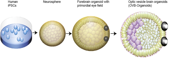
| The brain organoids grew symmetrical eye-like structures, which are those black blobs stuck to their bodies. |
 |
| How a brain organoid develops eyes. |
/https://tf-cmsv2-smithsonianmag-media.s3.amazonaws.com/filer/1e/51/1e51ce06-32aa-4957-90fc-8795663b5883/screen_shot_2021-08-19_at_111755_am.png) |
| The brain organoids grew symmetrical eye-like structures, which are those black blobs stuck to their bodies. |
 |
| How a brain organoid develops eyes. |
Mitochondria are the powerhouse of the cell, as any fifth-grade biology student could tell you. (Mitochondria is plural, one of them is called a mitochondrion). These tiny organelles turn the food, water, and air you consume into energy that can power your whole body. They also might be helping you see color more clearly.
Mitochondria are present in every cell, but not every cell gets the same amount. Generally, the more energy a cell needs, the more mitochondria it has. Your hardworking heart muscle cells, for example, are rich with them, having about 5,000 per cell! Your skin cells, by comparison, have just a few hundred.
Another cell type with a plenitude of mitochondria: your eyes. Specifically, inside your retinas, the light-sensitive tissue in the back of your eyeballs, exists a specialized type of cells called cone cells that allow us to see color. Cone cells are broadly organized into an outer segment, which picks up light, and an inner segment, which handles the rest of the cell's functions. It is in the inner segment that mitochondria cluster into a long bundle in inner segment of the cell.
 |
| Diagram of a cone cell |
Initially, it was thought that this glut of mitochondria produced energy for the cone cells. That, like heart muscle cells, they were using up a lot of energy. But researchers found that most of the energy produced in the cone cells came from glycolysis, a separate process that doesn't involve the mitochondria at all.
Evolutionarily, though, this doesn't make sense. There'd be no reason to pack so many mitochondria into cone cells if they were just sitting there. But if they weren't making energy, what were they doing?
The answer to this riddle came as a result of a rather morbid experiment. Scientists chose 13-lined ground squirrels as their model organism, since they're diurnal (coming out during the day and sleeping at night), and so have lots of cone cells for sensing color. The squirrels were raised in captivity for 5 months, fed cat chow and given some bedding and a PVC pipe for enrichment. On their day of reckoning they were gassed and decapitated by guillotine. (I am not making this up, it's in the "Methods" section of the research article. And I am still thinking about their little squirrel heads rolling)
 |
| They gave their lives for science. |
Their eyes were dissected, with the retinas cut into tiny pieces which were then fixed to microscope slides. Layers were peeled away until only the light-detecting cone cells remained, then a light was shined onto the live cells, mimicking the passage of light through the eyes. While those bundles of mitochondria might be expected to scatter the light, they instead focused it on the light-sensing outer segment of the cone cells. The oily membrane of the mitochondria had special reflective properties that made them "microlenses" for incoming light, creating a higher-resolution image.
While the study was carried out on squirrels, it has several implications for humans' eyes as well. For example, it helps explain the Stiles-Crawford effect, a phenomenon where color is perceived differently when seen through the pupil versus the edge of the eye. Through experiments and computer modeling, researchers saw that the mitochondrial interaction with light lined up with the Stiles-Crawford effect. In humans, this could be a useful way to diagnose eye disease, since many eye diseases cause mitochondrial dysfunction.
Here's a helpful explanation in the form of a video.