
These tiny blobs are called brain organoids, and are made by manipulating and growing stem cells. A stem cell, broadly speaking, is an undifferentiated cell, meaning it has the potential to turn into any other type of cell, being it bone, brain, or blood. When you were an embryo (in case you can't remember), you were made up of only stem cells, which divided and changed into all the different organs in your body. Scientists can use cultured stem cells in the same way, adding specific proteins that changes them into miniature livers, hearts, or, in this case, brains with light-sensitive eyes. They're basically blobs of brain tissue, incapable of thoughts or emotions, which makes them useful when employing real brains would be expensive, or at the least very ethically dubious.
/https://tf-cmsv2-smithsonianmag-media.s3.amazonaws.com/filer/1e/51/1e51ce06-32aa-4957-90fc-8795663b5883/screen_shot_2021-08-19_at_111755_am.png) |
| The brain organoids grew symmetrical eye-like structures, which are those black blobs stuck to their bodies. |
Like other organoids, growing miniature eyes in the lab has been done before, but not like this. Using stem cells, other researchers managed to create optic cups, the structures which go on to form almost the entire globe of the eye. However, in this experiment, researchers wanted to grow eye structures together with brain tissue to see how the two interacted as they developed together.
The results of the experiment are not unlike something out of science fiction. After adding retinol acetate, or vitamin A, to neural stem cells, eye cups developed within 30 days, and the visible, symmetrical eye structures we can see came in at about 50 days, mirroring the timeline of eye development in embryos. And, though they look like just black blobs, the rudimentary eyes contained different retinal cell types as well as lens and corneal tissue. They also formed connections to the rest of the neural tissue, and were sensitive to light, producing electrical impulses when exposed to it.
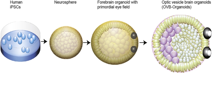 |
| How a brain organoid develops eyes. |
Being able to grow tiny brains with eyes isn't just cool and a bit frightening, though. Watching eyes as they develop can help us figure out what causes blindness at birth, or to analyze how different growth conditions might impact eye development and cause disease. They could even be cultured from a specific person's cells to create personalized transplants. Though these brain organoids can't think for themselves, they are giving us a lot to think about as far as the future of healing our eyes.


