Monday, April 25, 2022

Where Do (Eel) Babies Come From?

    Animal migrations are nothing sort of fascinating: the Arctic tern flies from the Arctic circle to the Antarctic, monarch butterflies travel the entire East Coast to end up in Mexico. Eels take a long journey, too: from the shores of Europe all the way to the Sargasso Sea, where they mate and give rise to their flat, clear-as-glass larvae. Or so we think. The truth is that no one has actually seen an eel mate in the wild.

    Even from ancient times the origins of eels have puzzled us; where and how they reproduce is still unknown. Aristotle thought that they wriggled up from the soil when it rained. In ancient Egypt it was believed they spawned from sunlight soaking the shores of the Nile. Freud dissected hundreds of eels trying to discover their sex organs; he was unsuccessful and refused to acknowledge the research. 
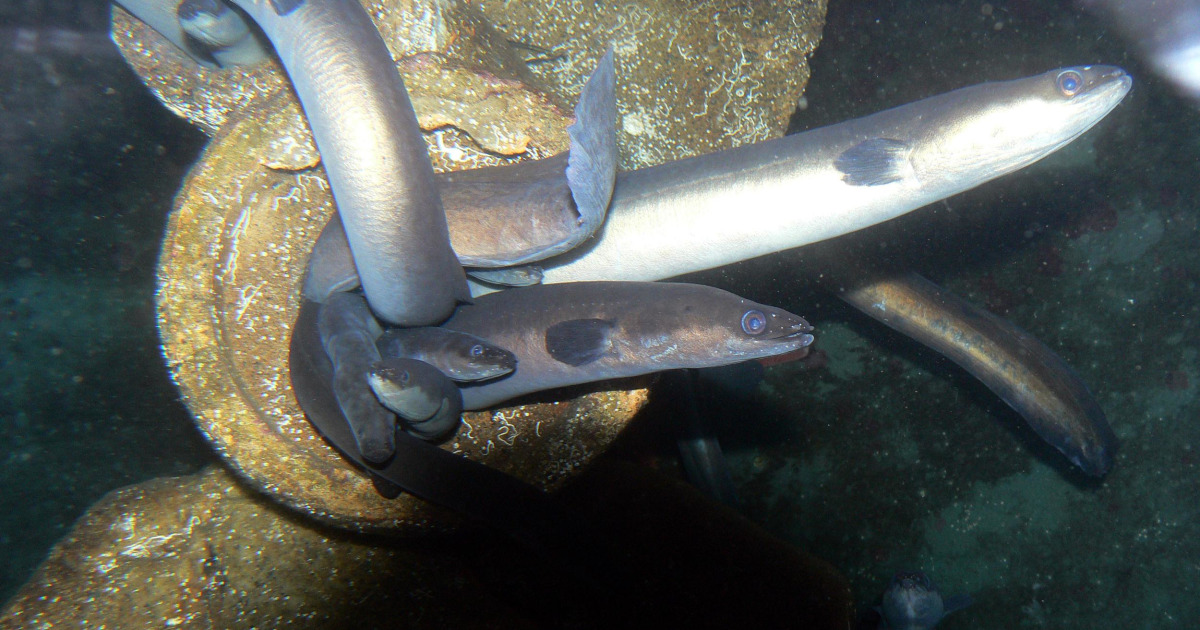
Maybe they're just a little shy.

    Today, we know that eels, specifically the European eel Anguilla anguilla, follow a predictable life pattern. They begin as flat, clear larvae, and mature into "glass eels", where they ride the Jet Stream to the coast of Europe. Once there, they migrate up the tiniest rivers and creeks, and then pull themselves onto land to colonize ponds and lakes. An eel on land can receive up to 50% of its oxygen from the air, making this journey less difficult than it sounds. Over the course of a decade, these young eels, or elvers, mature into the adult silver eels, and this is where their mating mystery begins.
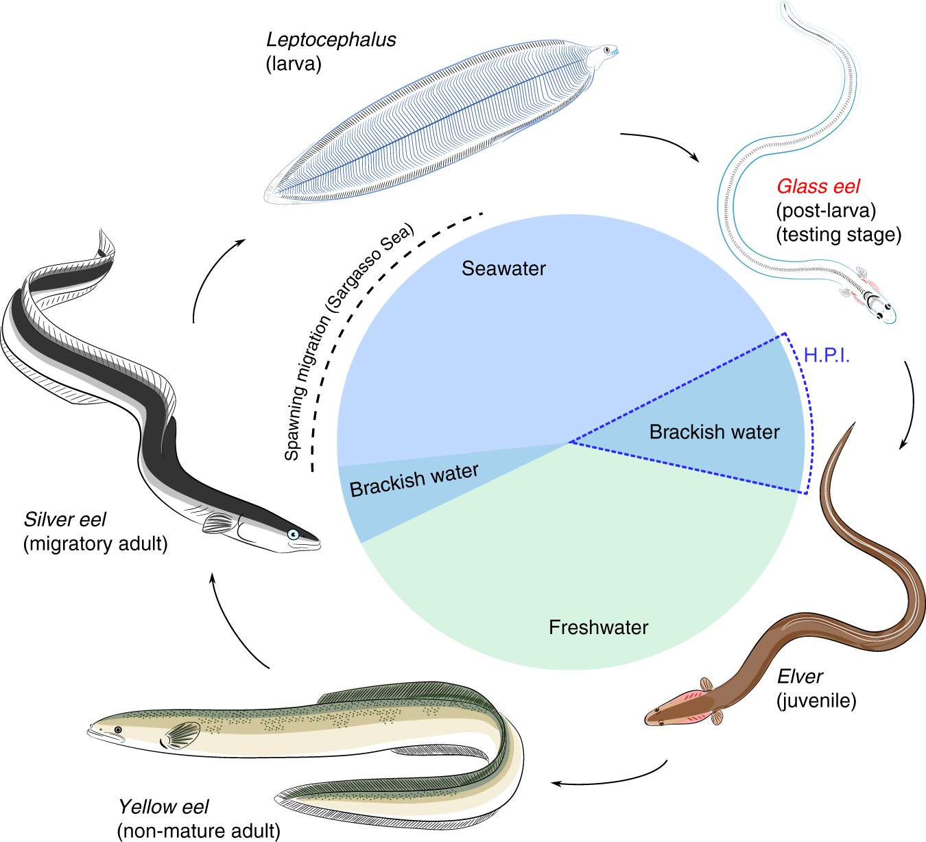
Eel life cycle. Leptocephalus larvae were once thought to be a completely separate species.

    In the 20th century, Danish biologist Johannes Schmidt set out to track down the eel's origins. By capturing juvenile eels, he compared their sizes to see where the smallest and youngest eels were coming from, eventually pinpointing them to the Sargasso, that calm, shoreless sea near the Caribbean. But, though we have an idea where they spawn, no one has actually seen them do it. Eels have successfully reproduced in captivity, and, unlike Freud, we have located their sex organs. But the finer points are missing: no eggs or young larvae have ever been found.

    A new hypothesis, published in 2020, aims to narrow down the area that European eels may be spawning in. With the Sargasso stretching 2 million square miles, the chances of two hot single eels meeting in the same area is slim (barring some kind of aquatic Tinder). Using virtual eel simulations based on earlier observations of the migrations, researchers proposed a new spawning hotspot along the mid-Atlantic ridge; more precisely, where this oceanic mountain range hits a salinity front, an area near the Azores islands, outside of the Sargasso. The cues of both the change in terrain and salinity could make this location much easier to find for mating eels. The Japanese eel, a related species, is known reliably to spawn at the intersection of a seamount ridge and a salinity front, adding credence to this theory. Manganese, a trace element associated with underwater volcanoes, was also found in high concentration in the bodies of European eels, but not in Sargasso sea larvae. 

"Green and magenta circles indicate areas where American and European eels have been observed, respectively8, and the white dotted box indicates the examined area for the numerical experiment."

    While eels were plentiful throughout Europe in the past, being a cheap food enjoyed by common people, today their populations have declined up to 90%, largely due to overfishing, parasites, and habitat loss. While it also solves a long-standing scientific mystery, discovering the mating habits of eels could also be a step towards keeping them alive.

Eels were once so common in Britain they were cooked in stock and allowed to gelatinize, creating the dish of jellied eels, popular throughout London among the poor. Or, why British people should never be allowed around food.  


Thursday, April 7, 2022

The Perfect Witness Can't Lie. But Can They Buzz?

Insects are everywhere in our lives: visiting our flowers, poking through our trash, or hiding out in our cupboards. Most would consider them disgusting, especially the flies and maggots that hang around rotten food. Rotten food, though, has a lot in common with rotting bodies, and flies aren't picky. Investigators of crime scenes can use these scavenging bugs, then, to tell the story that a victim can't. In the field of forensic entomology, insect evidence gives valuable clues about how and where a killing happened.

Forensic entomology as a field dates back as far as forensics itself. Collected Cases of Injustice Rectified, a book from 13th century China written by judge and scholar Song Ci, is the first ever book of forensic science, detailing how to identify causes of death and perform autopsies. It is also the first known application of forensic entomology. 

In 1235, Song Ci writes, a stabbing occurred in a Chinese village. The weapon of attack was determined to be a sickle after various sharp objects were tested against an animal corpse. All the villagers were ordered to place their sickles on the ground, but only one of them attracted blow flies, which were drawn to the remnants of blood, tissue, and hair left on it. The owner of the sickle confessed to the murder and was led away ashamed.

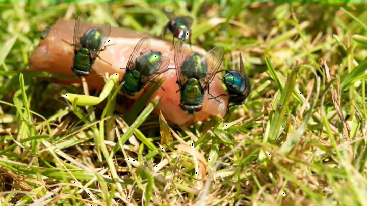
Blowflies on some dead stuff. It's hard to find pictures when you have a massive phobia of maggots. Ew ew ew.

After nearly 8 centuries, the field has become considerably more sophisticated. Insects collected from corpses can give valuable information about both the time and location of death, and even if drugs are present in the deceased's system. 

Time of death is the most easily approximated, using what we know about insect life cycles. First on the scene, and most valuable to investigators, are flies, usually blowflies or fleshflies. Attracted by the chemicals created by decomposition, these scavengers feed near orifices such as the mouth, or open wounds, blowflies colonizing the body within a few minutes of death. 

Later on, flies lay their eggs on the body, which hatch into maggots. By identifying the species and age of the maggots, investigators can find out how long ago they hatched, and approximate a time of death that way. Other insects come along during later stages of decomposition: beetles, mites, and moths. Entomological data can be incredibly accurate: before 24 hours, it is even more accurate than soft tissue analysis in determining time of death, and after 72 hours, insect evidence is sometimes the only method of determination.

One of the major concerns of forensic entomology is finding bibles small enough to swear in the witnesses.

The insects present can also tell where the death took place with surprising specificity. For example, if flies are on a corpse that aren't native to where it was found, this is good evidence that the body was moved after death. Insects in general also like warm, moist environments. If a body is left out in the sun, then, larvae will exhibit faster development, and be more numerous. The same is true if in a humid or rainy place, allowing investigators to pinpoint which areas are likely for a murder.

Even still, insects can be drug-tested for compounds present in the corpse when a toxicology report is impossible or needs more evidence, such as when soft tissues or bodily fluids have decayed. In the emerging field of entomotoxicology, investigators can test insects to find if substances are present in their tissue. Some substances also alter the time of development: generally, cocaine and methamphetamine cause faster growth, while some poisons, such as barbiturates, slow down development.

In an age where many forensic techniques are being proved unreliable or even pseudoscientific, new approaches to convicting killers must be based on hard evidence. Bite mark analysis, bullet lead comparison, and fingerprinting, all things we think are damning evidence, have actually been shown to be highly unreliable. Forensic entomology fortunately seems less "buggy", and as the field expands, we may see more flies and beetles convicting bad guys.

Thursday, March 31, 2022

A New Horrific State of Consciousness

The end is nigh! Scientists are growing brains in jars confined to a horrific existence: fully aware, but unable to move, to breathe, to experience, stripped of their ability to even cry out in despair. 
Well, not exactly. This experiment, which went viral on the internet for conjuring a very I Have No Mouth and I Must Scream flavor of terror, isn't as terrifying as you might glean from the tweet about it above. These little brains aren't conscious, have rudimentary structure, and are only the size of peas. Even so, growing brains and eyes from scratch gives us valuable information about how these organs develop in embryos, and how changes to that development leads to disease.

These tiny blobs are called brain organoids, and are made by manipulating and growing stem cells. A stem cell, broadly speaking, is an undifferentiated cell, meaning it has the potential to turn into any other type of cell, being it bone, brain, or blood. When you were an embryo (in case you can't remember), you were made up of only stem cells, which divided and changed into all the different organs in your body. Scientists can use cultured stem cells in the same way, adding specific proteins that changes them into miniature livers, hearts, or, in this case, brains with light-sensitive eyes. They're basically blobs of brain tissue, incapable of thoughts or emotions, which makes them useful when employing real brains would be expensive, or at the least very ethically dubious.
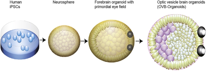
The brain organoids grew symmetrical eye-like structures, which are those black blobs stuck to their bodies.

Like other organoids, growing miniature eyes in the lab has been done before, but not like this. Using stem cells, other researchers managed to create optic cups, the structures which go on to form almost the entire globe of the eye. However, in this experiment, researchers wanted to grow eye structures together with brain tissue to see how the two interacted as they developed together.

The results of the experiment are not unlike something out of science fiction. After adding retinol acetate, or vitamin A, to neural stem cells, eye cups developed within 30 days, and the visible, symmetrical eye structures we can see came in at about 50 days, mirroring the timeline of eye development in embryos. And, though they look like just black blobs, the rudimentary eyes contained different retinal cell types as well as lens and corneal tissue. They also formed connections to the rest of the neural tissue, and were sensitive to light, producing electrical impulses when exposed to it.
How a brain organoid develops eyes.
Being able to grow tiny brains with eyes isn't just cool and a bit frightening, though. Watching eyes as they develop can help us figure out what causes blindness at birth, or to analyze how different growth conditions might impact eye development and cause disease. They could even be cultured from a specific person's cells to create personalized transplants. Though these brain organoids can't think for themselves, they are giving us a lot to think about as far as the future of healing our eyes.

Thursday, March 24, 2022

Mitochondria: Powerhouse of the Eyes?

Mitochondria are the powerhouse of the cell, as any fifth-grade biology student could tell you. (Mitochondria is plural, one of them is called a mitochondrion). These tiny organelles turn the food, water, and air you consume into energy that can power your whole body. They also might be helping you see color more clearly.

Mitochondria are present in every cell, but not every cell gets the same amount. Generally, the more energy a cell needs, the more mitochondria it has. Your hardworking heart muscle cells, for example, are rich with them, having about 5,000 per cell! Your skin cells, by comparison, have just a few hundred.

Another cell type with a plenitude of mitochondria: your eyes. Specifically, inside your retinas, the light-sensitive tissue in the back of your eyeballs, exists a specialized type of cells called cone cells that allow us to see color. Cone cells are broadly organized into an outer segment, which picks up light, and an inner segment, which handles the rest of the cell's functions. It is in the inner segment that mitochondria cluster into a long bundle in inner segment of the cell.

Diagram of a cone cell

Initially, it was thought that this glut of mitochondria produced energy for the cone cells. That, like heart muscle cells, they were using up a lot of energy. But researchers found that most of the energy produced in the cone cells came from glycolysis, a separate process that doesn't involve the mitochondria at all.

Evolutionarily, though, this doesn't make sense. There'd be no reason to pack so many mitochondria into cone cells if they were just sitting there. But if they weren't making energy, what were they doing?

The answer to this riddle came as a result of a rather morbid experiment. Scientists chose 13-lined ground squirrels as their model organism, since they're diurnal (coming out during the day and sleeping at night), and so have lots of cone cells for sensing color. The squirrels were raised in captivity for 5 months, fed cat chow and given some bedding and a PVC pipe for enrichment. On their day of reckoning they were gassed and decapitated by guillotine. (I am not making this up, it's in the "Methods" section of the research article. And I am still thinking about their little squirrel heads rolling)

They gave their lives for science.

Their eyes were dissected, with the retinas cut into tiny pieces which were then fixed to microscope slides. Layers were peeled away until only the light-detecting cone cells remained, then a light was shined onto the live cells, mimicking the passage of light through the eyes. While those bundles of mitochondria might be expected to scatter the light, they instead focused it on the light-sensing outer segment of the cone cells. The oily membrane of the mitochondria had special reflective properties that made them "microlenses" for incoming light, creating a higher-resolution image.

While the study was carried out on squirrels, it has several implications for humans' eyes as well. For example, it helps explain the Stiles-Crawford effect, a phenomenon where color is perceived differently when seen through the pupil versus the edge of the eye. Through experiments and computer modeling, researchers saw that the mitochondrial interaction with light lined up with the Stiles-Crawford effect. In humans, this could be a useful way to diagnose eye disease, since many eye diseases cause mitochondrial dysfunction.

Here's a helpful explanation in the form of a video.


Thursday, March 10, 2022

Ultrasound Could Save an Endangered Sea Snail

Ultrasound is one of the safest, easiest, and most useful methods of medical imaging available today. Ultrasound probes emit sound at a higher frequency than you can hear, which echoes off of tissues inside the body, returning to the probe to create an image. It's not unlike a bat's echolocation. Most people associate the technology with pregnancy. However, scientists at UC Davis are taking ultrasound out of the doctor's office and into the ocean to help recover one of our critically endangered sea creatures, the abalone.

Black Abalone, a critically endangered species on the IUCN Red List
Abalone make up several species of flat, spiral-shaped sea snails that live stuck to rocks in the shallow ocean, feeding on algae. They're prized for the iridescent mother-of-pearl that lines the insides of their shells, which is harvested to make jewelry or other decorations, and for their meat, which is a delicacy the world over. They're also major players in marine ecosystems, being food sources for marine mammals and helping to maintain kelp forests and reefs.

In recent years, though, their numbers have dwindled as a result of overfishing and ocean acidification, which erodes their shells with low pH. To aid in their survival and sustainability, scientists and farmers raise abalone in captivity, as in UC Davis's white abalone captive breeding program. However, telling when an abalone is about to spawn is difficult without being able to look inside at its gonads. And that's exactly what the researchers did.

To study an abalone requires prying it off of the rock it sticks to, a stressful experience that can harm the animal. Ultrasound technology, however, is far less invasive. To give an abalone an ultrasound, though, there's no jelly or gender reveal. The abalone is submerged in a tank, and the ultrasound wand is pressed to the outside of the tank, next to the abalone's foot. On a computer, the abalone's gonads show up as a thick dark band. The thicker the band, the more ready the abalone is to spawn. Identifying which snails are ready to spawn is useful for both abalone farmers and conservation experts to know which are going to be best for reproducing. For all the hardships abalone have faced, this is a promising step for reestablishing the creatures throughout the oceans.

Here's an interesting video of an ultrasound being given to an abalone.


Tuesday, March 8, 2022

Chocolate Frogs are Real in Peru

A previously unrecorded species discovered in the Peruvian Amazon recently has been going viral for its unusual appearance. Some liken it to a melted Tootsie roll, or to a chocolate frog straight out of Harry Potter. And check out that long nose! When new species go viral, it's usually because they have some fascinating characteristics either in looks or in how they impact our understanding of ecosystems. The Tapir Frog, while being small, has both.
Big long nose, like a tapir!


Quarter-sized, with a thick, blobby body ending in a distinctive pointed snout, the Tapir Frog is a striking creature, even if it spends most of its live hidden in soil and debris. That snout indicates that it probably spends most of its life nosing through the dirt. While most soil would be too hard and dense for frogs, who aren't known for being good diggers, the Tapir Frog has a rare and specific habitat where it thrives. The Amazon peatlands, damp areas where decaying plant matter litters the floor, make a perfect home for the tiny critters, who spend most of their lives underground, moving, eating, and laying their eggs, slipping in and out of the dense peat with their slick bodies.

Its discovery came as a result of a mass inventory survey in the Amazon, in a relatively untouched and unstudied area. Earlier that day scientists had discovered a tiny juvenile of the new species. Later, in the wee hours of the morning, they heard a strange peeping under the ground. After a frantic search, they dug up two adult male specimens, who had their genetic code analyzed to prove that they were indeed part of a new species. Germán Chávez, who spotted the frog along with a team of herpetologists (amphibian experts), shares in the sentiment that it "looks like it was made from chocolate."

While new to scientists, the Tapir Frog was already known by the local indigenous people of the Comunidad Nativa Tres Esquinas. When shown the frog, they identified it as "rana danta," or "Tapir Frog". Its scientific name is Synapturanus danta, the first name for the genus of frogs it's in, and the second being the local word for Tapir. One of its special abilities could be to be an indicator of Amazon peatland health: soil too dry would be uninhabitable for the little frogs. While only three specimens of this elusive frog were found and documented, scientists hope to uncover more to better understand the rich and rare ecosystem from which it comes.

Wednesday, February 23, 2022

A Feeling in the Pit of Your Stomach

Do you think your brain is a rational actor? That is, do you think with your "head" and not your "heart" or your "gut"? (This blog is going to become Jonathan Frakes Asks You Things, isn't it?) We have a tendency to attribute different parts of our cognition to parts of the body - the heart is emotional, the gut instinctive, the eyes superficial, the brain logical. However, these are merely metaphors, and as we all know, only the brain is responsible for processing information and making decisions. Is it really, though?

The digestive tract has its own nervous system, known as the enteric nervous system. The enteric nervous system is a division of the autonomic nervous system, which consists of those nerves that transmit information to and from the internal organs. Some other functions of the autonomic nervous system include regulating your heart rate and breathing, all unconscious processes, meaning they happen without you needing to manually control them. Imagine having to think about contracting your heart every second, and you'll be thankful you have this nervous system.

The enteric nervous system consists of all the nerves that control digestive functions, including peristalsis (the contraction of the intestines to move food along). Lining your digestive tract are over 200 million neurons, at least as many as in the spinal cord. Like your heartbeat or breathing, its functions are automatic, not consciously controlled. They even have the capacity to continue without the brain telling them to do so. When the vagus nerve, the main messenger from the brain to the autonomic nervous system, is severed, digestive functions were found to still continue.

There's a lot of layers going on here. Basically, nerves hide out between the muscles and the inner surface of the digestive tract.


There is some evidence and much speculation that the enteric nervous system could have an effect on cognitive processes, such as emotions and decision-making. While the enteric nervous system on a basic level creates the sensations of nausea and bloating, much of the information it sends is processed subconsciously, and can possibly determine mood. The enteric nervous system may also relay information from the pounds of microorganisms living in the gut, which can impact emotion and behavior. So your "gut feeling" could be a feeling from your gut after all.

Who is really holding the reins, then? New discoveries about the enteric nervous system challenge our brain-centric perception of behavior and mood. They can also change the way we diagnose, research, and treat certain neurological disorders, such as Autism Spectrum Disorder, Parkinson's, and ALS. Digestive dysfunctions are common along with the neurological symptoms of these disorders. With autism, a certain gene that is associated with the disorder is also associated with poor motility in the gut. These studies are developing, and mostly done on mice, but it remains to be seen how much of what we think of as brain dysfunction is closely linked to the gut.

Wednesday, February 16, 2022

The Heart-Throat and Your Cousin the Sea Squirt

Do you ever think about what it was like to be a single cell, eyeless and undifferentiated? Then, as you split into more and more cells, those parts of you migrated and took on different roles - light-sensing cells went to the eyes, cells lined themselves up in layers to make your three germ layers, and to divide you into a front side and a back side, a head and a tail. Your whole digestive system used to be just one big tube. 

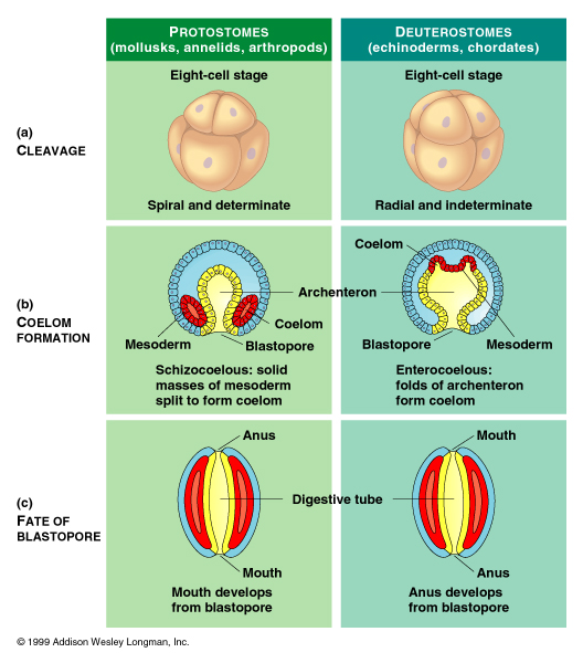
Arthropods and mollusks form mouth-first while chordates (like mammals) and echinoderms (sea stars, urchins) form anus-first. Sadly, I know some people who have never passed this stage of development.

It turns out that the way you develop can reveal a lot about your ancestry - what animals you're most closely related to, that is, and how they adapted to their environments, changing their body forms over time. 

For example, the origin of the heart still puzzles animal researchers. Its complex, multi-chambered structure in vertebrates isn't exactly mirrored by what was thought to be their closest relatives - the lancelet, a small, elongated, fish-like sea creature lacking jaws or sense organs. They have no distinct heart, just a blood vessel pumping colorless blood throughout the body. Enter the sea squirt, or tunicate - a little foreign-looking, but more related to chordates and more valuable to understanding the origin of human hearts.

Grandpa?

I know, it doesn't look like someone you'd see at a family reunion, but tunicates have complex body structures, including a U-shaped heart that squeezes blood through it. Sometimes, it actually reverses the direction of blood flow, though the reason for this isn't known. In their larval stage, they also have a notochord - a rod of cartilage that supports the body. However, they lose this when they mature and become sessile, or unmoving. The notochord in humans becomes the backbone.

Scientists researching cell differentiation in tunicates found that, when labeling cells that would form the heart, some of these cells wandered off to form the pharynx. This patch of cells, called "cardiopharyngeal", for how it gave rise to both the heart and muscles of the pharynx and jaw, is common to vertebrates and tunicates - similar differentiation has been observed in mice. The throat and the heart, organs from two very different systems, are linked, like childhood friends, based on what group of cells they came from. And vertebrates and tunicates are probably more linked than was previously thought. This also establishes how, in vertebrates, development of the respiratory, circulatory, and muscular systems are linked by ancient evolutionary trends, far back enough to connect you with your sea-squirt ancestry.

https://www.nature.com/articles/d41586-022-00413-y